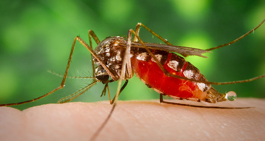
There are no teensy cups. But a urine test for wild mosquitoes has for the first time proved it can give an early warning that local pests are spreading diseases.
Mosquito traps remodeled with a pee-collecting card picked up telltale genetic traces of West Nile and two other worrisome viruses circulating in the wild, researchers in Australia report April 4 in the Journal of Medical Entomology.
The tests were based on an innovative saliva monitoring system unveiled in 2010: traps that lure mosquitoes into tasting honey-coated cards. Among its advantages, this card-based medical testing doesn’t need the constant refrigeration that checking whole mosquitoes does. And it’s not as labor intensive as monitoring sentinel chickens or pigs for signs of infection. But testing traces of mosquito saliva left on these cards comes close to the limits of current molecular methods for detecting viruses. In part, it’s an issue of volume. A mosquito drools fewer than five nanoliters of saliva when it tastes a card. In comparison, mosquitoes excrete about 1.5 microliters of liquid per pee, offering a veritable flood of material. So Dagmar Meyer of James Cook University in Cairns, Australia and her colleagues created urine collectors using standard overnight light traps and longer-standing traps that exhale delicious carbon dioxide, a mosquito come-hither.
The team set out 29 urine traps in two insect-rich spots in Queensland along with traps equipped to catch mosquito saliva. When mosquitoes fell for the trick and entered a urine trap, their excretions dripped through a mesh floor onto a collecting card. Adding a moist wick of water kept trapped mosquitoes alive and peeing longer, thus improving the sample. Pee traps picked up three viruses — West Nile, Ross River and Murray Valley encephalitis — while the saliva ones detected two, the researchers report.
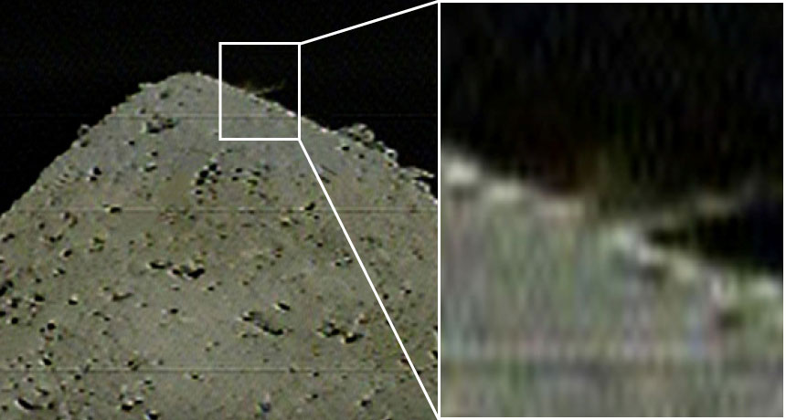
Hayabusa2 has blasted the asteroid Ryugu with a projectile, probably adding a crater to the small world’s surface and stirring up dust that scientists hope to snag.
The projectile, a two-kilogram copper cylinder, separated from the Hayabusa2 spacecraft at 9:56 p.m. EDT on April 4, JAXA, Japan’s space agency, reports.
Hayabusa2 flew to the other side of the asteroid to hide from debris that would have been ejected when the projectile hit (SN: 1/19/19, p. 20). Scientists won’t know for sure whether the object successfully made a crater, and, if so, how big it is, until the craft circles back. But by 10:36 p.m. EDT, Hayabusa2’s cameras had captured a blurry shot of a dust plume spurting up from Ryugu, so the team thinks the attempt worked. “This is the world’s first collision experiment with an asteroid!” JAXA tweeted.
Hayabusa2 plans to briefly touch down inside the crater to pick up a pinch of asteroid dust. The spacecraft has already grabbed one sample of Ryugu’s surface (SN Online: 2/22/19). But dust exposed by the impact will give researchers a look at the asteroid’s subsurface, which has not been exposed to sunlight or other types of space radiation for up to billions of years.
If all goes as planned, Hayabusa2 will return to Earth with both samples in late 2020. A third planned sample pickup has been scrapped because Ryugu’s boulder-strewn surface is so hazardous for the spacecraft. Comparing the two samples will reveal details of how being exposed to space changes the appearance and composition of rocky asteroids, and will help scientists figure out how Ryugu formed (SN Online: 3/20/19). Scientists hope that the asteroid contains water and organic material that might help explain how life got started in the solar system.
My youngest child, now just over a year old, has started to talk. Even though I’ve experienced this process with my older two, it’s absolutely thrilling. He is putting words to the thoughts that swirl around in his sweet little head, making his mind a little less mysterious to the rest of us.
But these early words may not mean what we think they mean, a new study hints. Unsurprisingly, when 2-year-olds were asked a series of “this or that” questions, the toddlers showed strong preferences — but not for the reasons you’d think. Overwhelmingly, the toddlers answered the questions with the last choice given. That bias, described in PLOS ONE on June 12, suggests that young children’s answers to these sorts of questions don’t actually reflect their desires. Instead, kids may simply be echoing the last thing they heard.
This verbal quirk can be used by parents to great effect, as the researchers point out in the title of their paper: “Cake or broccoli?” More fundamentally, the results raise questions about what sort of information a verbal answer actually pulls out of a young child’s mind. This murkiness is especially troublesome when it comes to questions whose answers call for adult action, such as: “Did you hit your sister on purpose or on accident?”
In the first series of experiments, researchers led by Emily Sumner at the University of California, Irvine, asked 24 1- and 2-year-olds a bunch of two-choice questions, some of which involved a polar bear named Rori or a grizzly bear named Quinn. One question, for example, was, “Does Rori live in an igloo or a tepee?” Later, the researchers switched the bear and the order of the options, asking, for example, “Does Quinn live in a tepee or an igloo?”
The toddlers could answer either verbally or, for reluctant speakers, by pointing at one of two stickers that showed the choices. When the children answered the questions by pointing, they chose the second option about half the time, right around chance. But when the toddlers spoke their answers, they chose the second option 85 percent of the time, regardless of the bear. SECOND BEST A toddler taking part in a study selects the second option in three either-or questions. This tendency, called the recency bias, may reflect kids’ inability to juggle several choices in their minds simultaneously. Credit: E. Sumner et al/PLOS ONE 2019
This abundance of second options selected — a habit known as the recency bias — might be due to the fact that young children have trouble holding the first option in mind, the researchers suspect. Other experiments showed that children’s tendency toward the second option got stronger when the words got longer.
Adults actually have the opposite tendency: We’re more inclined to choose the first option we’re given (the primacy bias). To see when this shift from last to first occurs, the researchers studied transcripts of conversations held between adults and children ages 1.5 to 4. In these natural conversations, 2-year-olds were more likely to choose the second option. But 3- and 4-year-olds didn’t show this bias, suggesting that the window closes around then.
The results hold a multitude of delightful parenting hacks: “Would you like to jump on the bed all night, or go to sleep?” But more importantly, the study serves as a reminder that the utterances of small children, while fascinating, may not carry the same meanings as those that come from more mature speakers. If you really want a straight answer, consider showing the two options to the toddler. But if you go that route, be prepared to hand over the cake.
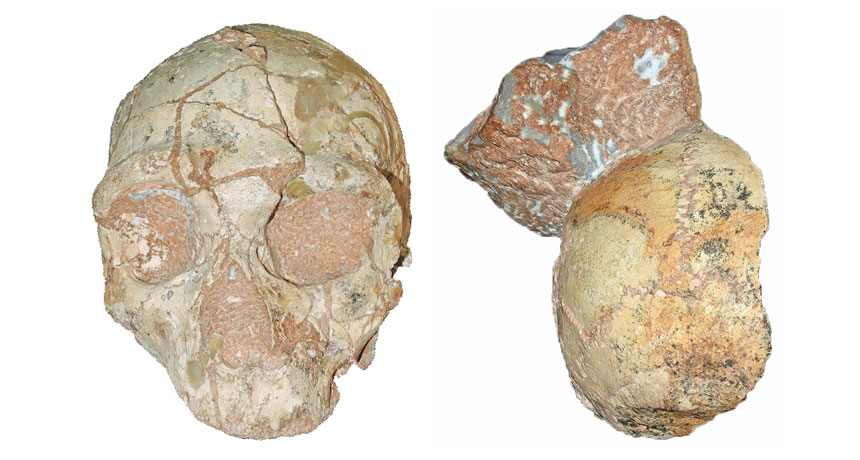
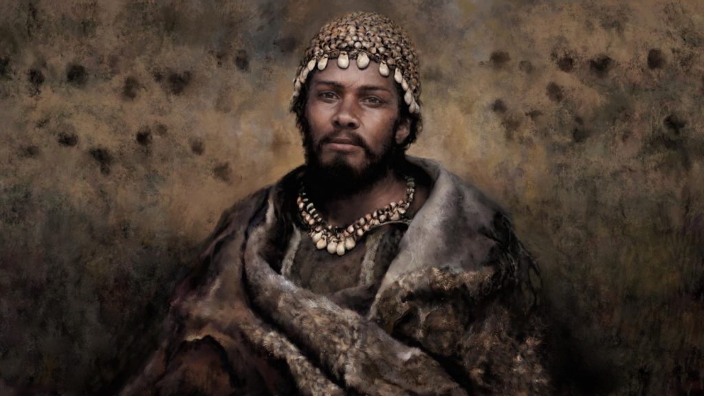
A skull found in a cliffside cave on Greece’s southern coast in 1978 represents the oldest Homo sapiens fossil outside Africa, scientists say.
That skull, from an individual who lived at least 210,000 years ago, was encased in rock that also held a Neandertal skull dating to at least 170,000 years ago, contends a team led by paleoanthropologist Katerina Harvati of the University of Tübingen in Germany.
If these findings, reported online July 10 in Nature, hold up, the ancient Greek H. sapiens skull is more than 160,000 years older than the next oldest European H. sapiens fossils (SN Online: 11/2/11). It’s also older than a proposed H. sapiens jaw found at Israel’s Misliya Cave that dates to between around 177,000 and 194,000 years ago (SN: 2/17/18, p. 6).
“Multiple Homo sapiens populations dispersed out of Africa starting much earlier, and reaching much farther into Europe, than previously thought,” Harvati said at a July 8 news conference. African H. sapiens originated roughly 300,000 years ago (SN: 7/8/17, p. 6). A small group of humans may have reached what’s now Greece more than 200,000 years ago, she suggested. Neandertals who settled in southeastern Europe not long after that may have replaced those first H. sapiens. Then humans arriving in Mediterranean Europe tens of thousands of years later would eventually have replaced resident Neandertals, who died out around 40,000 years ago (SN Online: 6/26/19).
But Harvati’s group can’t exclude the possibility that H. sapiens and Neandertals simultaneously inhabited southeastern Europe more than 200,000 years ago and sometimes interbred. A 2017 analysis of ancient and modern DNA concluded that humans likely mated with European Neandertals at that time.
The two skulls were held in a small section of wall that had washed into Greece’s Apidima Cave from higher cliff sediment and then solidified roughly 150,000 years ago. Since one skull is older than the other, each must originally have been deposited in different sediment layers before ending up about 30 centimeters apart on the cave wall, the researchers say. Earlier studies indicated that one Apidima skull, which retains the face and much of the braincase, was a Neandertal that lived at least 160,000 years ago. But fossilization and sediment pressures had distorted the skull’s shape. Based on four 3-D digital reconstructions of the specimen, Harvati’s team concluded that its heavy brow ridges, sloping face and other features resembled Neandertal skulls more than ancient and modern human skulls. An analysis of the decay rate of radioactive forms of uranium in skull bone fragments produced an age estimate of at least 170,000 years.
A second Apidima fossil, also dated using uranium analyses, consists of the back of a slightly distorted braincase. Its rounded shape in a digital reconstruction characterizes H. sapiens, not Neandertals, the researchers say. A bunlike bulge often protrudes from the back of Neandertals’ skulls. But without any facial remains to confirm the species identity of the partial braincase, “it is still possible that both Apidima skulls are Neandertals,” says paleoanthropologist Israel Hershkovitz of Tel Aviv University. Hershkovitz led the team that discovered the Misliya jaw and assigned it to H. sapiens.
Harvati and her colleagues will try to extract DNA and species-distinguishing proteins (SN: 6/8/19, p. 6) from the Greek skulls to determine their evolutionary identities and to look for signs of interbreeding between humans and Neandertals.
The find does little to resolve competing explanations of how ancient humans made their way out of Africa. Harvati’s suggestion that humans trekked from Africa to Eurasia several times starting more than 200,000 years ago is plausible, says paleoanthropologist Eric Delson of City University of New York’s Lehman College in an accompanying commentary. And the idea that some H. sapiens newcomers gave way to Neandertals probably also applied to humans who reached Misliya Cave and nearby Middle Eastern sites as late as around 90,000 years ago, before Neandertals occupied the area by 60,000 years ago, Delson says.
Hershkovitz disagrees. Ancient humans and Neandertals lived side-by-side in the Middle East for 100,000 years or more and occasionally interbred, he contends. Misliya Cave sediment bearing stone tools dates to as early as 274,000 years ago, Hershkovitz says. Since only H. sapiens remains have been found in the Israeli cave, ancient humans probably made those stone artifacts and could have been forerunners of Greek H. sapiens.
No one should have to sleep with the fishes, but new research on zebrafish suggests that we sleep like them.
Sleeping zebrafish have brain activity similar to both deep slow-wave sleep and rapid eye movement, or REM, sleep that’s found in mammals, researchers report July 10 in Nature. And the team may have tracked down the cells that kick off REM sleep.
The findings suggest that the basics of sleep evolved at least 450 million years ago in zebrafish ancestors, before the evolution of animals that give birth to live young instead of laying eggs. That’s 150 million years earlier than scientists thought when they discovered that lizards sleep like mammals and birds (SN: 5/28/16, p. 9).
What’s more, sleep may have evolved underwater, says Louis C. Leung, a neuroscientist at Stanford University School of Medicine. “These signatures [of sleep] really have important functions — even though we may not know what they are — that have survived hundreds of millions of years of evolution.” In mammals, birds and lizards, sleep has several stages characterized by specific electrical signals. During slow-wave sleep, the brain is mostly quiet except for synchronized waves of electrical activity. The heart rate decreases and muscles relax. During REM or paradoxical sleep, the brain lights up with activity almost like it’s awake. But the muscles are paralyzed (except for rapid twitching of the eyes) and the heart beats erratically.
For many years, scientists have known that fruit flies, nematodes, fish, octopuses and other creatures have rest periods reminiscent of sleep. But until now, no one could measure the electrical activity of those animals’ brains to see if that rest is the same as mammals’ snoozing.
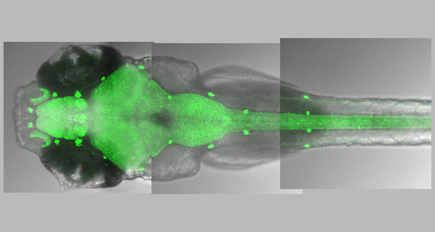
Leung and colleagues developed a system to do just that in zebrafish by genetically engineering them to make a fluorescent molecule that lights up when it encounters calcium, which is released when nerve cells and muscles are active. By following the flashes of light using a light sheet microscope, the researchers tracked brain and muscle activity in the naturally transparent fish larvae.
The next task was to lull fish asleep under the microscope. In some experiments, the team added drugs that trigger either slow-wave or REM sleep in mammals to the fish’s water. In others, researchers deprived fish of sleep for a night or tuckered the fish out with lots of activity during the day. Results from all the snooze-inducing methods were the same.
Sleeping fish have two distinct types of brain activity while sleeping, the team found. One, similar to slow-wave sleep, was characterized by short bursts of activity in some nerve cells in the brain. The researchers call that state slow-bursting sleep. REM-like sleep, which the researchers dubbed “propagating-wave sleep,” was characterized by frenzied brain activity that spreads like a wave through the brain. The researchers aren’t calling the sleep phases REM or slow-wave sleep because there are some minor differences between the way fish and mammals sleep. A group of cells that line hollow spaces called ventricles deep in the brain seems to trigger that wave of REM-like brain activity. These ependymal cells dip fingerlike cilia into the cerebral spinal fluid that bathes the ventricles and the central nervous system. The cells appear to beat their cilia faster as amounts of a well-known, sleep-promoting hormone called melanin-concentrating hormone in the fluid increases, the researchers discovered. It’s unclear how the ependymal cells communicate with the rest of the brain to set off REM-like activity. Such cells are also present in mammals, but no one has yet been able to see that deeply into the brains of sleeping mammals to determine whether the cells play a role in sleep. But knowing about these cells may help researchers develop better sleep aids, Leung says.
Just as in mammals, zebrafish’s whole bodies are affected during sleep. Their muscles relax during sleep and their hearts slow from about 200 beats per minute when awake to about 110 to 120 beats per minute while asleep during the slow-wave–like sleep. During the REM-like sleep, the heart slows even more to about 90 beats per minute and loses its regular rhythm. And the fish’s muscles also go completely slack. The one characteristic that the fish lack is rapid eye movement. Instead, the eyes roll back into their sockets, says study coauthor Philippe Mourrain, a biologist at Stanford University School of Medicine.
Lack of eye movement could indicate that emotion-processing parts of the brain, such as the amygdala, aren’t as active in zebrafish as they are in mammals, says sleep researcher Allan Pack of the University of Pennsylvania Perelman School of Medicine. With their brain-activity monitoring, the researchers have taken sleep research “to the next level,” says Pack, and “they present pretty compelling evidence” of slow-wave and REM-like sleep in the fish.
The whole-body involvement that the researchers documented solidifies the argument that fish sleep is similar to mammals, says neuroscientist Paul Shaw of Washington University School of Medicine in St. Louis. In all organisms known to snooze, “sleep is manifest everywhere” in the body, he says.
Future experiments may show why poor sleep or a lack of Zs contributes to health problems in people, such as obesity, heart disease and diabetes.
Ice sheets expanded across much of northern Europe from around 25,000 to 19,000 years ago, making a huge expanse of land unlivable. That harsh event set in motion a previously unrecognized tale of two human populations that played out at opposite ends of the continent.
Western European hunter-gatherers outlasted the icy blast in the past. Easterners got replaced by migrations of newcomers.
That’s the implication of the largest study to date of ancient Europeans’ DNA, covering a period before, during and after what’s known as the Last Glacial Maximum, paleogeneticist Cosimo Posth and colleagues report March 1 in Nature. As researchers have long thought, southwestern Europe provided refuge from the last Ice Age’s big chill for hunter-gatherers based in and near that region, the scientists say. But it turns out that southeastern Europe, where Italy is now located, did not offer lasting respite from the cold for nearby groups, as previously assumed.
Instead, those people were replaced by genetically distinct hunter-gatherers who presumably had lived just to the east along the Balkan Peninsula. Those people, who carried ancestry from parts of southwestern Asia, began trekking into what’s now northern Italy by about 17,000 years ago, as the Ice Age began to wane.
“If local [Ice Age] populations in Italy did not survive and were replaced by groups from the Balkans, this completely changes our interpretation of the archaeological record,” says Posth, of the University of Tübingen in Germany.
Posth and colleagues’ conclusions rest on analyses of DNA from 356 ancient hunter-gatherers, including new molecular evidence for 116 individuals from 14 countries in Europe and Asia. Excavated human remains that yielded DNA dated to between about 45,000 and 5,000 years ago (SN: 4/7/21).
Comparisons of sets of gene variants inherited by these hunter-gatherers from common ancestors enabled the researchers to reconstruct population movements and replacements that shaped ancient Europeans’ genetic makeup. For the first time, ancient DNA evidence included individuals from what’s known as the Gravettian culture, which dates from about 33,000 to 26,000 years ago in central and southern Europe, and from southwestern Europe’s Solutrean culture, which dates to between about 24,000 and 19,000 years ago. Contrary to expectations, makers of Gravettian tools came from two genetically distinct groups that populated western and eastern Europe for roughly 10,000 years before the Ice Age reached its peak, Posth says. Researchers have traditionally regarded Gravettian implements as products of a biologically uniform population that occupied much of Europe.
“What we previously thought was one genetic ancestry in Europe turned out to be two,” says paleogeneticist Mateja Hajdinjak of the Max Planck Institute for Evolutionary Anthropology in Leipzig, Germany, who did not participate in the new study. And “it seems that western and southwestern Europe served as a [refuge from glaciation] more than southeastern Europe and Italy.”
Descendants of the western Gravettian population, who are associated with Solutrean artifacts and remnants of another ancient culture in western Europe that ran from about 19,000 to 14,000 years ago, outlasted the Ice Age before spreading northeastward across Europe, the researchers say.
Further support for southwestern Europe as an Ice Age refuge comes from DNA extracted from a pair of fossil teeth that belonged to an individual linked to the Solutrean culture in southern Spain. That roughly 23,000-year-old adult was genetically similar to western European hunter-gatherers who lived before and after the Last Glacial Maximum, Max Planck paleogeneticist Vanessa Villalba-Mouco and colleagues, including Posth, report March 1 in Nature Ecology & Evolution.
Meanwhile, the genetic evidence suggests that hunter-gatherers in what’s now Italy were replaced by people from farther east, probably based in the Balkan region. Those newcomers must have brought with them a distinctive brand of stone artifacts, previously excavated at Italian sites and elsewhere in eastern Europe, known as Epigravettian tools, Posth says. Many archaeologists have suspected that Epigravettian items were products of hunter-gatherers who clustered in Italy during the Ice Age’s peak freeze.
But, Hajdinjak says, analyses of DNA from fossils of Ice Age Balkan people are needed to clarify what groups moved through Italy, and when those migrations occurred.
Ultimately, descendants of Ice Age migrants into Italy reached southern Italy and then western Europe by around 14,000 years ago, Posth and colleagues say. Ancient DNA evidence indicates that, during those travels, they left a major genetic mark on hunter-gatherers across Europe.
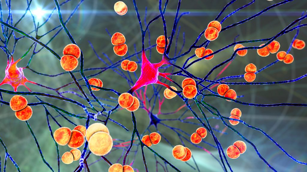
Bacteria can slip into the brain by commandeering cells in the brain’s protective layers, a new study finds. The results hint at how a deadly infection called bacterial meningitis takes hold.
In mice infected with meningitis-causing bacteria, the microbes exploit previously unknown communication between pain-sensing nerve cells and immune cells to slip by the brain’s defenses, researchers report March 1 in Nature. The results also hint at a new way to possibly delay the invasion — using migraine medicines to interrupt those cell-to-cell conversations. Bacterial meningitis is an infection of the protective layers, or meninges, of the brain that affects 2.5 million people globally per year. It can cause severe headaches and sometimes lasting neurological injury or death.
“Unexpectedly, pain fibers are actually hijacked by the bacteria as they’re trying to invade the brain,” says Isaac Chiu, an immunologist at Harvard Medical School in Boston. Normally, one might expect pain to be a warning system for us to shut down the bacteria in some way, he says. “We found the opposite…. This [pain] signal is being used by the bacteria for an advantage.”
It’s known that pain-sensing neurons and immune cells coexist in the meninges, particularly in the outermost layer called the dura mater (SN: 11/11/20). So to see what role the pain and immune cells play in bacterial meningitis, Chiu’s team infected mice with two of the bacteria known to cause the infection in humans: Streptococcus pneumoniae and S. agalactiae. The researchers then observed where that bacteria ended up in mice genetically tweaked to lack pain-sensing nerve cells and compared those resting spots to those in mice with the nerve cells intact.
Mice without pain-sensing neurons had fewer bacteria in the meninges and brain than those with the nerve cells, the team found. This contradicts the idea that pain in meningitis serves as a warning signal to the body’s immune system, mobilizing it for action.
Further tests showed that the bacteria triggered a chain of immune-suppressing events, starting with the microbes secreting toxins in the dura mater.
The toxins hitched onto the pain neurons, which in turn released a molecule called CGRP. This molecule is already known to bind to a receptor on immune cells, where it helps control the dura mater’s immune responses. Injecting infected mice with more CGRP lowered the number of dural immune cells and helped the infection along, the researchers found.
The team also looked more closely at the receptor that CGRP binds to. In infected mice bred without the receptor, fewer bacteria made it into the brain. But in ones with the receptor, immune cells that would otherwise engulf bacteria and recruit reinforcements were disabled. The findings suggest that either preventing the release of CGRP or preventing it from binding to immune cells might help delay infection.
In humans, neuroscientists know that CGRP is a driver of headaches — it’s already a target of migraine medications (SN: 6/5/18). So the researchers gave five mice the migraine medication olcegepant, which blocks CGRP’s effects, and infected them with S. pneumoniae. After infection, the medicated mice had less bacteria in the meninges and brain, took longer to show symptoms, didn’t lose as much weight and survived longer than mice that were not given the medication.
The finding suggests olcegepant slowed the infection. Even though it only bought mice a few extra hours, that’s crucial in meningitis, which can develop just as quickly. Were olcegepant to work the same way in humans, it might give doctors more time to treat meningitis. But the effect is probably not as dramatic in people, cautions Michael Wilson, a neurologist at the University of California, San Francisco who wasn’t involved with the work.
Scientists still need to determine whether pain-sensing nerve cells and immune cells have the same rapport in human dura mater, and whether migraine drugs could help treat bacterial meningitis in people.
Neurologist Avindra Nath has doubts. Clinicians think the immune response and inflammation damage the brain during meningitis, says Nath, who heads the team investigating nervous system infections at the National Institute of Neurological Disorders and Stroke in Bethesda, Md. So treatment involves drugs that suppress the immune response, rather than enhance it as migraine medications might.
Chiu acknowledges this but notes there might be room for both approaches. If dural mater immune cells could head the infection off at the pass, it may keep some bacteria from penetrating the defenses, minimizing brain inflammation.
This study might not ultimately change how clinicians treat patients, Wilson says. But it still reveals something new about one of the first lines of defense for the brain.