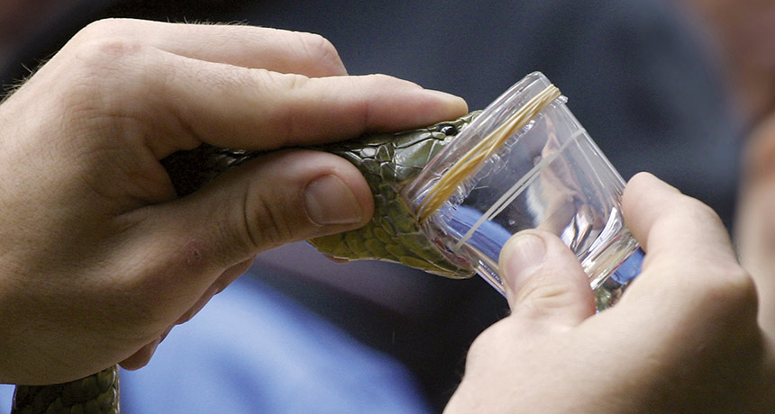
SAN DIEGO — Labs growing replicas of snakes’ venom glands may one day replace snake farms.
Researchers in the Netherlands have succeeded in growing mimics of venom-producing glands from multiple species of snakes. Stem cell biologist Hans Clevers of the Hubrecht Institute in Utrecht, the Netherlands, reported the creation of these organoids on December 10 at a joint meeting of the American Society for Cell Biology and the European Molecular Biology Organization.
If scientists can extract venom from the lab-grown glands, that venom might be used to create new drugs and antidotes for bites including from snakes that aren’t currently raised on farms.
Up to 2.7 million people worldwide are estimated to be bitten by venomous snakes each year. Between about 81,000 to 138,000 people die as a result of the bite, and as many as roughly 400,000 may lose limbs or have other disabilities, according to the World Health Organization. Antivenoms are made using venom collected from snakes usually raised on farms. Venom is injected into other animals that make antibodies to the toxins. Purified versions of those antibodies can help a bitten person recover, but must be specific to the species of snake that made the bite. “If it’s a fairly rare or local snake, chances are there would be no antidote,” Clevers says.
Three postdoctoral researchers in Clevers’ lab wanted to know if they could make organoids — tissues grown from stem cells to have properties of the organs they mimic — from snakes and other nonmammalian species. The researchers started with Cape coral snakes (Aspidelaps lubricus) that were dissected from eggs just before hatching. Stem cells taken from the unhatched snakes grew into several different types of organoids, including some that make venom closely resembling the snake’s normal venom, Clevers reported at the meeting.
His team has produced venom-gland organoids from at least seven species of snakes. The organoids have survived in the lab for up to two years so far.
Clevers and colleagues hope to harvest venom from the organoids, which produce more highly concentrated venom than snakes usually make. “It’s probably going to be easier than milking a snake,” he says.
Satellites may be a more accurate way to track smog-producing ammonia.
It’s notoriously tricky to pinpoint accurate numbers for ammonia gas emissions from sources such as animal feedlots and fertilizer plants. But new maps, generated from infrared radiation measurements gathered by satellites, reveal global ammonia hot spots in greater detail than before. The new data suggest that previous estimates underestimate the magnitude of these emissions, researchers report December 5 in Nature.
In the atmosphere, ammonia, which contains nitrogen, can help form tiny particles that worsen air quality and harm human health. The research could help keep tabs on who’s emitting how much, to make sure that factories and farms are meeting environmental standards. Emissions are usually estimated by adding up output from individual known sources of activity, but those calculations are only as good as the data that go into them. Ammonia sticks around only hours to a few days in the atmosphere, so on-the-ground measurements vary a lot even in the same place, says coauthor Martin Van Damme, an atmospheric scientist at the Université Libre de Bruxelles in Belgium.
“There’s so much uncertainty in ammonia emissions,” says Daven Henze, a mechanical engineer at the University of Colorado Boulder who wasn’t part of the research. Other scientists, including his research group, have estimated ammonia releases using satellite data before. But these new maps rely on a more detailed dataset and have substantially better resolution, Henze says — fine enough that the study authors were able to link areas of high emissions to specific factories or farms. The new maps show 248 nitrogen emission hot spots across the globe at a resolution of about a kilometer. Eighty-three of those hot spots arose from agricultural activity that involved high numbers of cows, pigs and chickens, such as a site in Colorado that overlapped on satellite imagery maps with two big cattle feedlots. Ammonia emissions from feedlots come largely from livestock waste. Another 158 sites were affected by industrial emissions — mostly from sites that produced ammonia-based fertilizer, such as in Marvdasht, Iran. Six hot spots couldn’t be pinned to specific activity. Ammonia is also emitted naturally, from volcanoes or seabird colonies. But most of those sources were too weak or not concentrated enough to show up as hot spots in the data. Lake Natron in Tanzania is the one exception — its mud flats show up as an ammonia-releasing hot spot, perhaps due to decaying algae. But it’s not clear why other lakes with similar mud flats didn’t. Some natural sources may have gone undetected because of where they were located — in places with heavy cloud cover that obscured the data, or where turbulent air dissipated ammonia especially quickly, Van Damme suggests.
Some areas with particularly high overall ammonia emissions from biomass burning or fertilizer, such as West Africa and the Indus Valley in Pakistan and northern India, didn’t reveal specific hot spots, either, the researchers report.
Since people in the United States began dying in the fentanyl-related drug overdose epidemic, whites have been hit the hardest. But new data released March 21 by the Centers for Disease Control and Prevention show that African-Americans and Hispanics are catching up.
Non-Hispanic whites still experience the majority of deaths involving fentanyl, a synthetic opioid. But among African-Americans and Hispanics, death rates rose faster from 2011 to 2016. Whites experienced a 61 percent annual increase, on average, while the rate rose 140.6 percent annually for blacks and 118.3 percent per year for Hispanics. No reliable data were available for other racial groups. Overall, the number of U.S. fentanyl-related deaths in 2011 and 2012 hovered just above 1,600. A sharp increase began in 2013, reaching 18,335 deaths in 2016. That’s up from 0.5 deaths per 100,000 people in 2011 to 5.9 per 100,000 in 2016.
In the first three years of the data, men and women died from fentanyl-related overdoses at similar rates, around 0.5 per 100,000. But in 2013, those paths diverged, and by 2016, the death rate among men was 8.6 per 100,000; for women it was 3.1 per 100,000. Overdose death rates rose most sharply along the East Coast, including in New England and the middle Atlantic, and in the Great Lakes region.
One of the most powerful opioids, fentanyl has been around for decades and is still prescribed to fight pain. But it has emerged as a street drug that is cheap to make and is found mixed into other drugs. In 2013, fentanyl was the ninth most common drug involved in overdose deaths, according to the CDC report; in 2016, it was number one. Just a little bit can do a lot of damage: The drug can quickly kill a person by overwhelming several systems in the body (SN: 9/3/2016, p. 14).
Viruses, which cannot reproduce on their own, infect cells and usurp their genetic machinery for use in making new viruses…. But just how viruses use the cell machinery is unknown.… Some answers may come from work with an unusual virus, called M13, that has a particularly compatible relationship with … [E. coli] bacteria. — Science News, April 5, 1969
Update M13 did help unlock secrets of viral replication. Some bacteria-infecting viruses, called bacteriophages or simply phages, kill the host cell after hijacking the cell’s machinery to make copies of themselves. Other phages, including M13, leave the cell intact. Scientists are using phage replication to develop drugs and technologies, such as virus-powered batteries (SN: 4/25/09, p. 12). Adding genetic instructions to phage DNA for making certain molecules lets some phages produce antibodies against diseases such as lupus and cancer. The technique, called phage display, garnered an American-British duo the 2018 Nobel Prize in chemistry (SN: 10/27/18, p. 16).
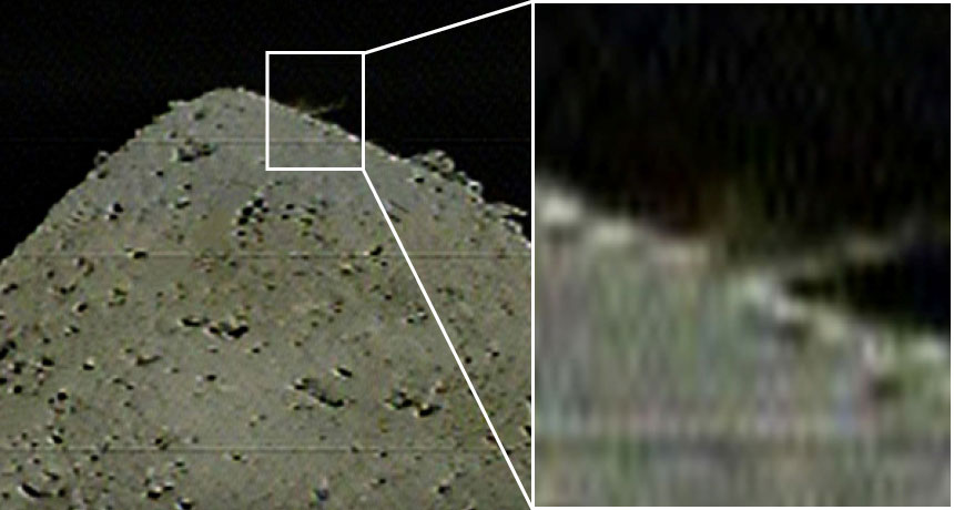
Hayabusa2 has blasted the asteroid Ryugu with a projectile, probably adding a crater to the small world’s surface and stirring up dust that scientists hope to snag.
The projectile, a two-kilogram copper cylinder, separated from the Hayabusa2 spacecraft at 9:56 p.m. EDT on April 4, JAXA, Japan’s space agency, reports.
Hayabusa2 flew to the other side of the asteroid to hide from debris that would have been ejected when the projectile hit (SN: 1/19/19, p. 20). Scientists won’t know for sure whether the object successfully made a crater, and, if so, how big it is, until the craft circles back. But by 10:36 p.m. EDT, Hayabusa2’s cameras had captured a blurry shot of a dust plume spurting up from Ryugu, so the team thinks the attempt worked. “This is the world’s first collision experiment with an asteroid!” JAXA tweeted.
Hayabusa2 plans to briefly touch down inside the crater to pick up a pinch of asteroid dust. The spacecraft has already grabbed one sample of Ryugu’s surface (SN Online: 2/22/19). But dust exposed by the impact will give researchers a look at the asteroid’s subsurface, which has not been exposed to sunlight or other types of space radiation for up to billions of years.
If all goes as planned, Hayabusa2 will return to Earth with both samples in late 2020. A third planned sample pickup has been scrapped because Ryugu’s boulder-strewn surface is so hazardous for the spacecraft. Comparing the two samples will reveal details of how being exposed to space changes the appearance and composition of rocky asteroids, and will help scientists figure out how Ryugu formed (SN Online: 3/20/19). Scientists hope that the asteroid contains water and organic material that might help explain how life got started in the solar system.
My youngest child, now just over a year old, has started to talk. Even though I’ve experienced this process with my older two, it’s absolutely thrilling. He is putting words to the thoughts that swirl around in his sweet little head, making his mind a little less mysterious to the rest of us.
But these early words may not mean what we think they mean, a new study hints. Unsurprisingly, when 2-year-olds were asked a series of “this or that” questions, the toddlers showed strong preferences — but not for the reasons you’d think. Overwhelmingly, the toddlers answered the questions with the last choice given. That bias, described in PLOS ONE on June 12, suggests that young children’s answers to these sorts of questions don’t actually reflect their desires. Instead, kids may simply be echoing the last thing they heard.
This verbal quirk can be used by parents to great effect, as the researchers point out in the title of their paper: “Cake or broccoli?” More fundamentally, the results raise questions about what sort of information a verbal answer actually pulls out of a young child’s mind. This murkiness is especially troublesome when it comes to questions whose answers call for adult action, such as: “Did you hit your sister on purpose or on accident?”
In the first series of experiments, researchers led by Emily Sumner at the University of California, Irvine, asked 24 1- and 2-year-olds a bunch of two-choice questions, some of which involved a polar bear named Rori or a grizzly bear named Quinn. One question, for example, was, “Does Rori live in an igloo or a tepee?” Later, the researchers switched the bear and the order of the options, asking, for example, “Does Quinn live in a tepee or an igloo?”
The toddlers could answer either verbally or, for reluctant speakers, by pointing at one of two stickers that showed the choices. When the children answered the questions by pointing, they chose the second option about half the time, right around chance. But when the toddlers spoke their answers, they chose the second option 85 percent of the time, regardless of the bear. SECOND BEST A toddler taking part in a study selects the second option in three either-or questions. This tendency, called the recency bias, may reflect kids’ inability to juggle several choices in their minds simultaneously. Credit: E. Sumner et al/PLOS ONE 2019
This abundance of second options selected — a habit known as the recency bias — might be due to the fact that young children have trouble holding the first option in mind, the researchers suspect. Other experiments showed that children’s tendency toward the second option got stronger when the words got longer.
Adults actually have the opposite tendency: We’re more inclined to choose the first option we’re given (the primacy bias). To see when this shift from last to first occurs, the researchers studied transcripts of conversations held between adults and children ages 1.5 to 4. In these natural conversations, 2-year-olds were more likely to choose the second option. But 3- and 4-year-olds didn’t show this bias, suggesting that the window closes around then.
The results hold a multitude of delightful parenting hacks: “Would you like to jump on the bed all night, or go to sleep?” But more importantly, the study serves as a reminder that the utterances of small children, while fascinating, may not carry the same meanings as those that come from more mature speakers. If you really want a straight answer, consider showing the two options to the toddler. But if you go that route, be prepared to hand over the cake.
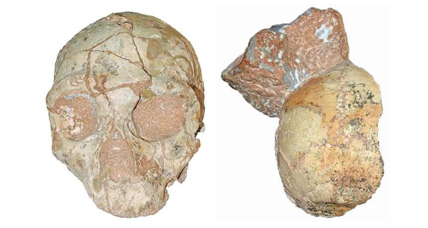
A skull found in a cliffside cave on Greece’s southern coast in 1978 represents the oldest Homo sapiens fossil outside Africa, scientists say.
That skull, from an individual who lived at least 210,000 years ago, was encased in rock that also held a Neandertal skull dating to at least 170,000 years ago, contends a team led by paleoanthropologist Katerina Harvati of the University of Tübingen in Germany.
If these findings, reported online July 10 in Nature, hold up, the ancient Greek H. sapiens skull is more than 160,000 years older than the next oldest European H. sapiens fossils (SN Online: 11/2/11). It’s also older than a proposed H. sapiens jaw found at Israel’s Misliya Cave that dates to between around 177,000 and 194,000 years ago (SN: 2/17/18, p. 6).
“Multiple Homo sapiens populations dispersed out of Africa starting much earlier, and reaching much farther into Europe, than previously thought,” Harvati said at a July 8 news conference. African H. sapiens originated roughly 300,000 years ago (SN: 7/8/17, p. 6). A small group of humans may have reached what’s now Greece more than 200,000 years ago, she suggested. Neandertals who settled in southeastern Europe not long after that may have replaced those first H. sapiens. Then humans arriving in Mediterranean Europe tens of thousands of years later would eventually have replaced resident Neandertals, who died out around 40,000 years ago (SN Online: 6/26/19).
But Harvati’s group can’t exclude the possibility that H. sapiens and Neandertals simultaneously inhabited southeastern Europe more than 200,000 years ago and sometimes interbred. A 2017 analysis of ancient and modern DNA concluded that humans likely mated with European Neandertals at that time.
The two skulls were held in a small section of wall that had washed into Greece’s Apidima Cave from higher cliff sediment and then solidified roughly 150,000 years ago. Since one skull is older than the other, each must originally have been deposited in different sediment layers before ending up about 30 centimeters apart on the cave wall, the researchers say. Earlier studies indicated that one Apidima skull, which retains the face and much of the braincase, was a Neandertal that lived at least 160,000 years ago. But fossilization and sediment pressures had distorted the skull’s shape. Based on four 3-D digital reconstructions of the specimen, Harvati’s team concluded that its heavy brow ridges, sloping face and other features resembled Neandertal skulls more than ancient and modern human skulls. An analysis of the decay rate of radioactive forms of uranium in skull bone fragments produced an age estimate of at least 170,000 years.
A second Apidima fossil, also dated using uranium analyses, consists of the back of a slightly distorted braincase. Its rounded shape in a digital reconstruction characterizes H. sapiens, not Neandertals, the researchers say. A bunlike bulge often protrudes from the back of Neandertals’ skulls. But without any facial remains to confirm the species identity of the partial braincase, “it is still possible that both Apidima skulls are Neandertals,” says paleoanthropologist Israel Hershkovitz of Tel Aviv University. Hershkovitz led the team that discovered the Misliya jaw and assigned it to H. sapiens.
Harvati and her colleagues will try to extract DNA and species-distinguishing proteins (SN: 6/8/19, p. 6) from the Greek skulls to determine their evolutionary identities and to look for signs of interbreeding between humans and Neandertals.
The find does little to resolve competing explanations of how ancient humans made their way out of Africa. Harvati’s suggestion that humans trekked from Africa to Eurasia several times starting more than 200,000 years ago is plausible, says paleoanthropologist Eric Delson of City University of New York’s Lehman College in an accompanying commentary. And the idea that some H. sapiens newcomers gave way to Neandertals probably also applied to humans who reached Misliya Cave and nearby Middle Eastern sites as late as around 90,000 years ago, before Neandertals occupied the area by 60,000 years ago, Delson says.
Hershkovitz disagrees. Ancient humans and Neandertals lived side-by-side in the Middle East for 100,000 years or more and occasionally interbred, he contends. Misliya Cave sediment bearing stone tools dates to as early as 274,000 years ago, Hershkovitz says. Since only H. sapiens remains have been found in the Israeli cave, ancient humans probably made those stone artifacts and could have been forerunners of Greek H. sapiens.
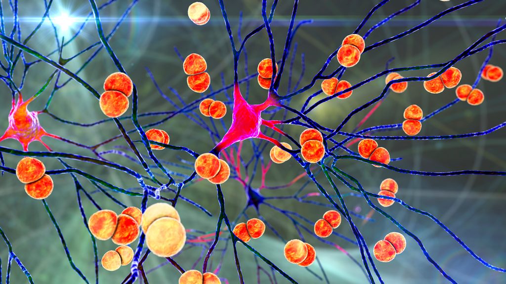
Bacteria can slip into the brain by commandeering cells in the brain’s protective layers, a new study finds. The results hint at how a deadly infection called bacterial meningitis takes hold.
In mice infected with meningitis-causing bacteria, the microbes exploit previously unknown communication between pain-sensing nerve cells and immune cells to slip by the brain’s defenses, researchers report March 1 in Nature. The results also hint at a new way to possibly delay the invasion — using migraine medicines to interrupt those cell-to-cell conversations. Bacterial meningitis is an infection of the protective layers, or meninges, of the brain that affects 2.5 million people globally per year. It can cause severe headaches and sometimes lasting neurological injury or death.
“Unexpectedly, pain fibers are actually hijacked by the bacteria as they’re trying to invade the brain,” says Isaac Chiu, an immunologist at Harvard Medical School in Boston. Normally, one might expect pain to be a warning system for us to shut down the bacteria in some way, he says. “We found the opposite…. This [pain] signal is being used by the bacteria for an advantage.”
It’s known that pain-sensing neurons and immune cells coexist in the meninges, particularly in the outermost layer called the dura mater (SN: 11/11/20). So to see what role the pain and immune cells play in bacterial meningitis, Chiu’s team infected mice with two of the bacteria known to cause the infection in humans: Streptococcus pneumoniae and S. agalactiae. The researchers then observed where that bacteria ended up in mice genetically tweaked to lack pain-sensing nerve cells and compared those resting spots to those in mice with the nerve cells intact.
Mice without pain-sensing neurons had fewer bacteria in the meninges and brain than those with the nerve cells, the team found. This contradicts the idea that pain in meningitis serves as a warning signal to the body’s immune system, mobilizing it for action.
Further tests showed that the bacteria triggered a chain of immune-suppressing events, starting with the microbes secreting toxins in the dura mater.
The toxins hitched onto the pain neurons, which in turn released a molecule called CGRP. This molecule is already known to bind to a receptor on immune cells, where it helps control the dura mater’s immune responses. Injecting infected mice with more CGRP lowered the number of dural immune cells and helped the infection along, the researchers found.
The team also looked more closely at the receptor that CGRP binds to. In infected mice bred without the receptor, fewer bacteria made it into the brain. But in ones with the receptor, immune cells that would otherwise engulf bacteria and recruit reinforcements were disabled. The findings suggest that either preventing the release of CGRP or preventing it from binding to immune cells might help delay infection.
In humans, neuroscientists know that CGRP is a driver of headaches — it’s already a target of migraine medications (SN: 6/5/18). So the researchers gave five mice the migraine medication olcegepant, which blocks CGRP’s effects, and infected them with S. pneumoniae. After infection, the medicated mice had less bacteria in the meninges and brain, took longer to show symptoms, didn’t lose as much weight and survived longer than mice that were not given the medication.
The finding suggests olcegepant slowed the infection. Even though it only bought mice a few extra hours, that’s crucial in meningitis, which can develop just as quickly. Were olcegepant to work the same way in humans, it might give doctors more time to treat meningitis. But the effect is probably not as dramatic in people, cautions Michael Wilson, a neurologist at the University of California, San Francisco who wasn’t involved with the work.
Scientists still need to determine whether pain-sensing nerve cells and immune cells have the same rapport in human dura mater, and whether migraine drugs could help treat bacterial meningitis in people.
Neurologist Avindra Nath has doubts. Clinicians think the immune response and inflammation damage the brain during meningitis, says Nath, who heads the team investigating nervous system infections at the National Institute of Neurological Disorders and Stroke in Bethesda, Md. So treatment involves drugs that suppress the immune response, rather than enhance it as migraine medications might.
Chiu acknowledges this but notes there might be room for both approaches. If dural mater immune cells could head the infection off at the pass, it may keep some bacteria from penetrating the defenses, minimizing brain inflammation.
This study might not ultimately change how clinicians treat patients, Wilson says. But it still reveals something new about one of the first lines of defense for the brain.
The Milky Way is churning out far more stars than previously thought, according to a new estimate of its star formation rate.
Gamma rays from aluminum-26, a radioactive isotope that arises primarily from massive stars, reveal that the Milky Way converts four to eight solar masses of interstellar gas and dust into new stars each year, researchers report in work submitted to arXiv.org on January 24. That range is two to four times the conventional estimate and corresponds to an annual birthrate in our galaxy of about 10 to 20 stars, because most stars are less massive than the sun. At this rate, every million years — a blink of the eye in astronomical terms — our galaxy spawns 10 million to 20 million new stars. That’s enough to fill roughly 10,000 star clusters like the beautiful Pleiades cluster in the constellation Taurus. In contrast, many galaxies, including most of the ones that orbit the Milky Way, make no new stars at all.
“The star formation rate is very important to understand for galaxy evolution,” says Thomas Siegert, an astrophysicist at the University of Würzburg in Germany. The more stars a galaxy makes, the faster it enriches itself with oxygen, iron and the other elements that stars create. Those elements then alter star-making gas clouds and can change the relative number of large and small stars that the gas clouds form.
Siegert and his colleagues studied the observed intensity and spatial distribution of emission from aluminum-26 in our galaxy. A massive star creates this isotope during both life and death. During its life, the star blows the aluminum into space via a strong wind. If the star explodes when it dies, the resulting supernova forges more. The isotope, with a half-life of 700,000 years, decays and gives off gamma rays.
Like X-rays, and unlike visible light, gamma rays penetrate the dust that cloaks the youngest stars. “We’re looking through the entire galaxy,” Siegert says. “We’re not X-raying it; here we’re gamma-raying it.”
The more stars our galaxy spawns, the more gamma rays emerge. The best match with the observations, the researchers find, is a star formation rate of four to eight solar masses a year. That is much higher than the standard estimate for the Milky Way of about two solar masses a year.
The revised rate is very realistic, says Pavel Kroupa, an astronomer at the University of Bonn in Germany who was not involved in the work. “I’m very impressed by the detailed modeling of how they account for the star formation process,” he says. “It’s a very beautiful work. I can see some ways of improving it, but this is really a major step in the absolutely correct direction.”
Siegert cautions that it is difficult to tell how far the gamma rays have traveled before reaching us. In particular, if some of the observed emission arises nearby — within just a few hundred light-years of us — then the galaxy has less aluminum-26 than the researchers have calculated, which means the star formation rate is on the lower side of the new estimate. Still, he says it’s unlikely to be as low as the standard two solar masses per year. In any event, the Milky Way is the most vigorous star creator in a collection of more than 100 nearby galaxies called the Local Group. The largest Local Group galaxy, Andromeda, converts only a fraction of a solar mass of gas and dust into new stars a year. Among Local Group galaxies, the Milky Way ranks second in size, but its high star formation rate means that we definitely try a lot harder.
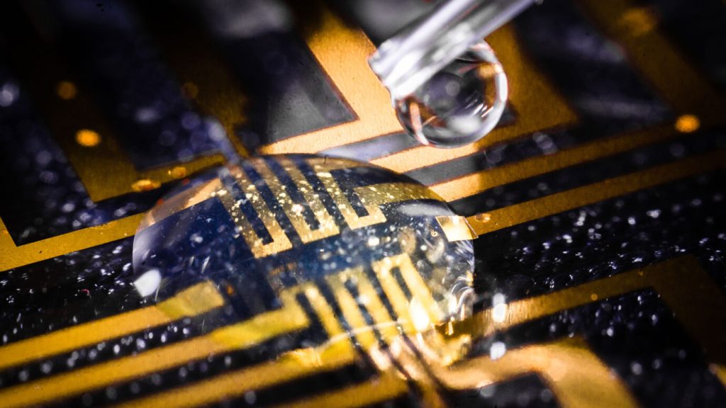
For the first time, researchers have harnessed the body’s own chemistry to “grow” electrodes inside the tissues of living fish, blurring the boundary between biology and machines.
The technique uses the body’s sugars to turn an injected gel into a flexible electrode without damaging tissues, experiments show. Zebrafish with these electrodes grown in their brains, hearts and tail fins showed no signs of ill effects, and ones tested in leeches successfully stimulated a nerve, researchers report in the Feb. 24 Science. Someday, these electrodes could be useful for applications ranging from studying how biological systems work to improving human-machine interfaces. They also could be used in “bioelectronic medicine,” such as brain stimulation therapies for depression, Parkinson’s disease and other conditions (SN: 2/10/19).
Soft electronics aim to bridge the gap between soft, curvy biology and electronic hardware. But these electronics typically still must carry certain parts that can be prone to cracks and other issues, and inserting these devices inevitably causes damage to tissues.
“All the devices we have made, even though we have made them flexible, to make them more soft, when we introduce them, there will still be a scar. It’s like sticking a knife into the organ,” says Magnus Berggren, a materials scientist at Linköping University in Sweden. That scarring and inflammation can degrade electrode performance over time.
Previous efforts to grow soft electronics inside tissues have drawbacks. One approach uses electrical or chemical signals to power the transformation from chemical soup to conducting electrodes, but these zaps also cause damage. A 2020 study got around this problem by genetically modifying cells in worms to produce an engineered enzyme that does the job, but the new method achieves its results without genetic alterations.
Berggren and colleagues’ electrodes instead exploit a natural energy source already present in the body: sugars. The gel cocktail contains molecules called oxidases that react with the sugars — glucose or lactate — to produce hydrogen peroxide. That then activates another ingredient in the cocktail, an enzyme called hydrogen peroxidase, which is the catalyst needed to transform the gel into a conducting electrode.
“The approach leverages elegant chemistry to overcome many of the technical challenges,” says biomedical engineer Christopher Bettinger of Carnegie Mellon University in Pittsburgh, who was not involved in the study.
To test the technique, the researchers injected the cocktail into the brains, hearts and tail fins of transparent zebrafish. The gel turns blue when it becomes conductive, giving a visual readout of its success. “The beautiful thing is you can see it: The zebrafishes’ tail changes color, and we know that blue indicates a conducting polymer,” says materials scientist Xenofon Strakosas, also of Linköping University. “The first time I saw it, I thought ‘Wow, it’s really working!’”
The fish appeared to suffer no ill effects, and the researchers saw no evidence of tissue damage. In partially dissected leeches, the team showed that delivering a current to a nerve via a soft electrode could induce muscle contractions. Ultimately, devices like this could be paired with various wireless technologies in development.
Long-term implant performance remains to be determined, however. “The demonstrations are impressive,” Bettinger says. “What remains to be seen is the stability of the electrode.” Over time, substances in the body could react with the electrode materials, degrading it or even producing toxic substances.
The team still needs to refine how precisely the electrodes can stimulate nerves, says chemical engineer Zhenan Bao of Stanford University, who was not involved in the work. She and colleagues developed the way to “grow” electrical components using genetic modifications. Ensuring stimulation is concentrated where it’s needed for a treatment, while preventing the leakage of current to unwanted regions will be important, she says.
In the new study, the relative abundance of different sugars in different tissues determines exactly where electrodes form. But in the future, a component of the main ingredient could be swapped out for elements that attach to specific bits of biology to make targeting much more precise, Berggren says. “We’re conducting experiments right now where we’re trying to bind these materials directly on individual cells.” Notes Strakosas: “There are some applications where precision is really important; that’s where we have to invest effort.”